Ultraviolet RTI
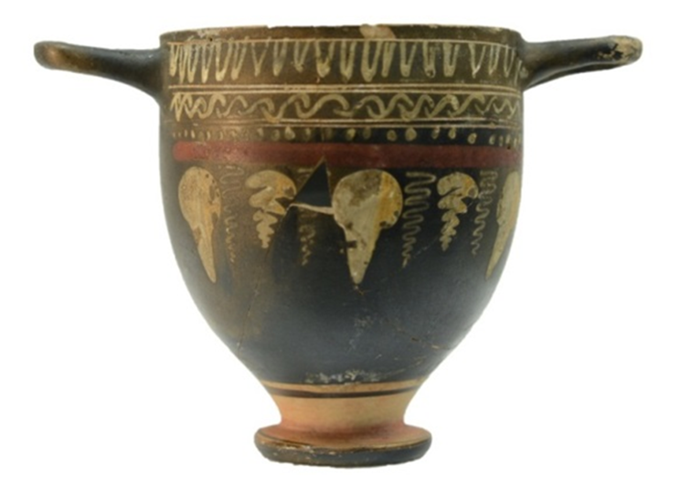
A painted and incised ceramic vessel was used as case study in an attempt to evaluate the efficiency of Reflected UV (UVR) RTI and UV induced visible fluorescence (UVF) RTI.
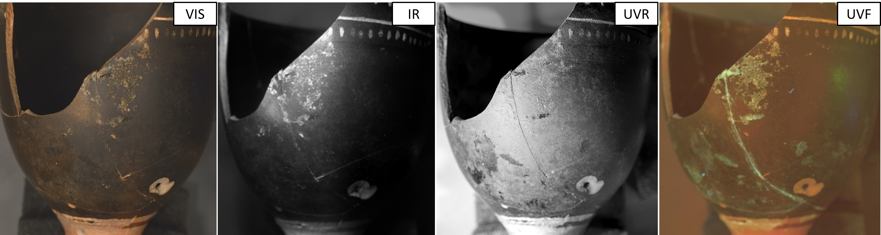
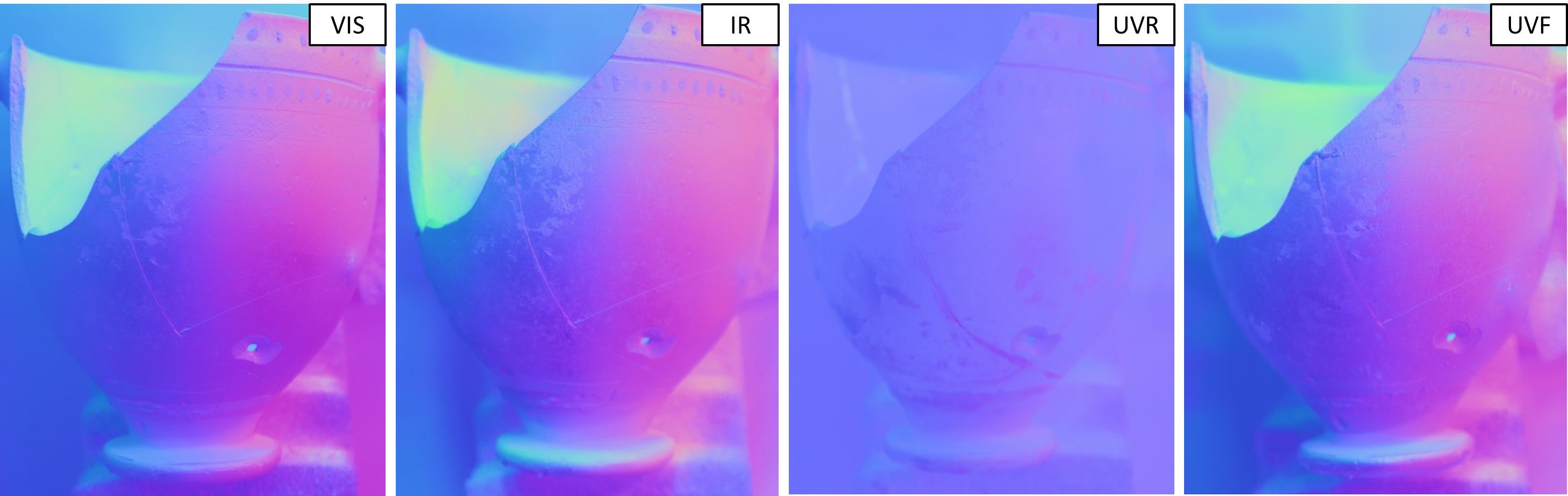
The Highlight RTI data capture took place at the archaeological imaging laboratory of the University of Southampton, using a UV-VIS-IR modified DSLR camera, adequate filters and lighting. The reflected UV-RTI datasets were captured with a UV transmitter and an IR barrier filter. The UV induced visible fluorescence RTI datasets were captured with an IR and an UV barrier filter.
The UV induced visible fluorescence image reveals the remains of conservation materials on the surface of the ceramic skyphos and the previous repairs, because of the visible fluorescence emission of common adhesives used. The UV induced visible fluorescence RTI enables the user to virtually move the radiation source around the object leading to numerous different visualizations. In that way the conservator can reach a better understanding about the morphology of the previous repairs.
Reflected UV-RTI offers the opportunity for enhanced examination of subtle surface variations; the remains of the conservation materials, the differentiations of the glaze due to its poor preservation and the salts’ efflorescence. The technique provided in a single file a combination of axial and UV imaging. It is notable that the axial positioning of radiation sources in reflected UV imaging is advantageous and is proposed for recording of scratches and smudges.
The synergy of RTI and UV imaging, results in an enhanced methodology for non-destructive examination and leads to different views of artefacts, which can be used for documentation, presentation, communication and research purposes.
UV-RTI visualization:
- Emphasizes subtle variations in the outer layer that are not clear in the visible spectral area
- Reveals episodes of the museum life of the artefact
- Reveals manufacture evidence and decay relevant to varnish layer or glazes
Leave a Reply